Identifying middle ear disorders can often be a challenging task, simply because all relevant structures are hidden behind the surface of the ear drum. At Acoustic Insight, we are developing Aurisvue: an easy-to-use, handheld scope that enables physicians to investigate the middle ear beyond the surface of the ear drum in real time! Find out more about our innovation below.
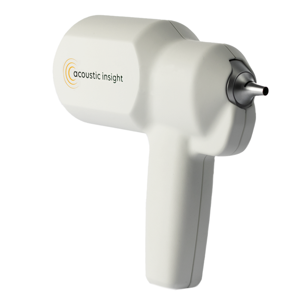
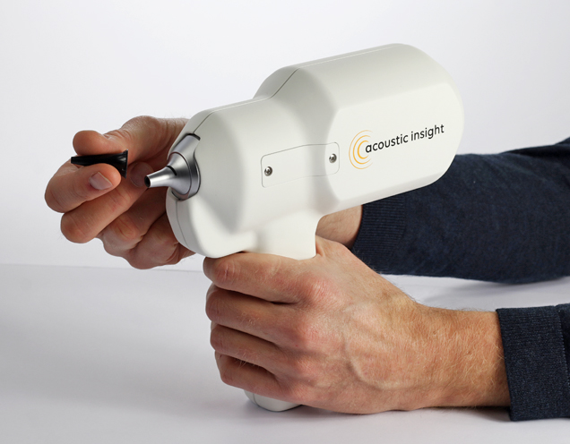
Aurisvue is designed to fit seamlessly into the standard clinical workflow. Our handheld probe operates in a similar manner as conventional otoscopes that is intuitive for clinicians. A built-in color camera provides live feedback during the examination. Aurisvue supports ear specula with different diameters and fits easily into a typical exam room due to its small physical footprint.
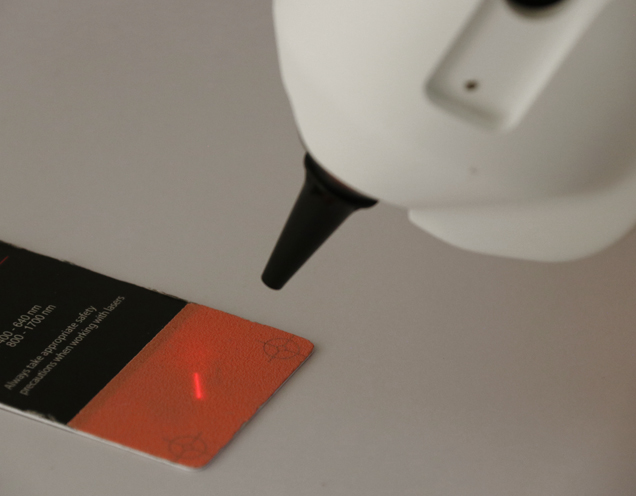
OCT is a noninvasive and non-contact imaging technique that uses infrared laser light to capture micrometer resolution images in depth. Our handheld scope directs the scanning laser beam to the surface of the ear drum and displays echo-like images of the tympanic membrane and middle ear space on the computer screen – in real-time! Aurisvue has been designed to maximize transmission through the ear drum, cover the entire depth of the middle ear cavity while maintaining micrometer scale resolution.
Aurisvue produces volumetric images of the middle ear space in just a few seconds. Clinicians can scroll through hundreds of densely spaced slices providing detailed information about the shape, structure and dimensions of the tympanic membrane, ossicular chain and other middle ear structures. The volumetric images can also be used to measure distances between the tympanic membrane and hearing bones.

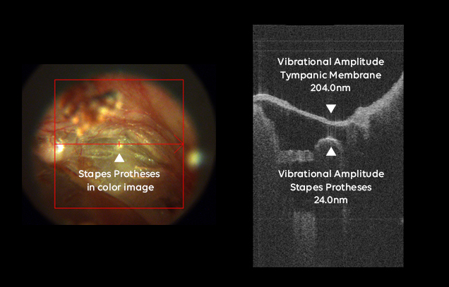
Aurisvue is also capable of measuring nanometer scale vibrations of the tympanic membrane and hearing bones. A sound stimulus incorporated in our handheld probe can emit pure tones in the frequency range of 500Hz to 20.000Hz to stimulate movement of the entire ossicular chain. The ability to measure these nanometer-scale amplitudes of each hearing bone separately is unique compared to the existing standard of care.